Educational:
Systematize factual knowledge about the structure of cells of plants and animals, prokaryotes and eukaryotes, about the functions of the main organelles of the cell, nucleus, membranes;
Show the unity of all life on earth based on knowledge of cell theory;
To form the concept of a cell as an open biological system, a structural and functional unit of life on earth;
Show the relationship (integration) of cytological knowledge with the development of painting, music, and historically.
Educational:
Cultivate interest in the subject of biology, cultivate self-demandingness, perseverance, and artistic qualities.
Lesson of generalization and systematization of knowledge.
Equipment:
- Tables “Structure of a plant cell”, “Structure of a living cell”. Music by Vivaldi, Tchaikovsky. Still lifes of Dutch painters. Microscopes, teaching material, multimedia projector, microscope, microslides.
I
– Class organization.
II
– Setting the goals and objectives of the lesson.
Teacher:
Since the 6th grade we have known that cytology is the science of the cell, and the cell is the structural and functional unit of life on Earth. We learned a lot about the complex structure of the cell, the history of its discovery and the creation of cell theory.
In today's lesson we must summarize our work, summarize the materials, briefly recall the history of the development of cells and the creation of cell theory, prove that the cell is the structural unit of life on Earth, repeat the structure of cell organelles, compare plant and animal cells, prokaryotes and eukaryotes. Write down the topic of our lesson: “Generalization of knowledge on the topic “cell”.”
Cell. Who is she? You and I think about each of our lessons. A small lump of mucus that can only be seen through a microscope or the smallest particle of the Universe in which all the concepts of life fit? To better understand this, let's return mentally and visually to those distant times when people first uttered the word “cell”. And the guys from one of the creative groups who take an active part in preparing for the lesson and have artistic abilities will help us with this.
Student:
The 17th century is a century of stagnation and discovery, contradictions and upswings in science, art, and literature.
Teacher:
The 17th century was a time when people turned to nature, seeing in it the origins of life. Nature is in everything: in music, painting, literature. You hear this miraculous music of the great composer Vivaldi, who lived and worked at this time. The “Seasons” sound, and this is the sound of a spring stream and the chirping of birds in the blue sky. On the board are works of Dutch landscape painters of the 17th century and here you see beautiful pictures of nature.
In the same century, a whole tribe of progressive naturalists appeared who tried to penetrate the most intimate secrets of nature. Who can you name? What events occurred in biology that revolutionized views on the structure of living beings?
Student:
1665 - English physicist Robert Hooke (1653 - 1703), examining a section of a cork under a microscope, discovered a very interesting phenomenon, and what exactly...
Teacher:
Let's take a look together. How it was?
Student:
I am very grateful to this Italian Galileo Galilei, who created an instrument called a “microscope”, which helped me see something of great interest to the whole world. We, microscopists of our time, stand on the threshold of great things and great discoveries. I worked a lot, parried a lot, and everywhere: on the core of an elderberry, on the stem of a reed, on the cork of any other tree, under a microscope I saw coeluses (cells), cells that were lined up in more or less dense rows in my field of vision! Oh, miracle! Oh, the beauty and eternal harmony of nature!
Student:
1680 - Dutch nature explorer Antonie van Leeuwenhoek discovers animal cells - protozoa.
Student:
The research of many microscopists around the world is taking a rather interesting turn, and I think my discoveries will also lead to something. This drop of stagnant water that is standing in my yard has long since turned green. But what can I see in her? (He leans over the microscope without enthusiasm.) Oh eureka! People, what do I see! In this small drop of dirty water I met a whole world of small living creatures. A world that is immediately difficult to understand, comprehend, and explain.
Teacher:
Much time has passed since then, the 17th century died down, was replaced by the 18th, then the 19th century. This time in science was filled with new discoveries, facts and contradictions. The Italian Malpighi, the Englishman Gruchech Purkine, the Germans Schleiden and Schwani contributed to the history of the study of the cell.
By the 30s of the 19th century, a lot of information, facts, and new knowledge about the cell had accumulated, which needed to be systematized and generalized. Nature, the concept of life, is always beautiful, either in the works of great composers or in the discoveries of great biologists. The science of the 19th century, which is capable of summarizing the facts accumulated over the past centuries and explaining discoveries, does not stand still. Who will do this?
Student:
These are the outstanding figures of 19th century cytology, Schleiden and Schwann.
Student:
It’s already 1839, a famous year in my life, here is the work created over many years of my life (reads) “Microscopic studies of the structure and growth of animals and plants.” The well-known botanist Schleiden helped me a lot, I am very grateful to him! By collaborating with him, this is what we came up with. I will read to you, respectable citizens, very interesting postulates from my book.
Postulate No. 1: “All organisms consist of essentially identical parts; it is from these cells that they are formed and grow according to essentially identical laws.”
Postulate No. 2: “Each cell within certain boundaries is an individual, independent whole. But these individuals act together in such a way that a harmonious whole emerges. All tissues are composed of cells or are formed from cells.”
Student:
Let me read out the third postulate of our theory, which is considered very important.
“The general principle of development for the elementary particles of the body is cell formation. At first there is a structureless non-living substance, yes, yes, that is exactly it, which lies between the cells. Cells are formed from this substance according to certain laws.”
Student:
Yes, this is a true creation of science, this is the cellular theory.
Student:
Just a moment, my friends! Yes, these are not entirely accurate postulates! Let me introduce you, Virchow is a German pathologist, please love and favor him! And anyone who has read my book “Cellular Pathology” will understand that the third postulate of Schleiden and Schwann is imperfect and that it will be replaced by a new one from my creation.
“Every cell comes from another cell. Where a cell arises, a cell must have preceded it, just as an animal comes from an animal, a plant from a plant.”
Teacher:
This is how the history of the development of society goes, from victories to defeats, from failure to ascent. This story is complex, the path to understanding science is thorny. But the moment of discovery is always beautiful, just like this music that sounds today!
Cell organelles
Cell organelles are specialized cell structures that perform various vital functions. The cells of protozoa are especially complex, where one cell makes up the entire organism and performs the functions of respiration, excretion, digestion and many others.
Cell organelles are divided into:
- Non-membrane - ribosomes, cell center, microtubules, movement organelles (flagella, cilia)
- Single-membrane - ER, Golgi complex (apparatus), lysosomes and vacuoles
- Double membrane - nucleus, plastids, mitochondria
Before talking about the organelles of a cell, without which its vital activity is impossible, it is necessary to mention something without which a cell does not exist at all - the cell membrane. The cell membrane limits the cell from the outside world and forms its internal environment.
Cell membrane (shell)
Remember that unlike the cell wall, which only plant cells and fungal cells have (it gives them a dense, rigid shape), all cells without exception have a cell membrane! I will explain this important point once again. Animal cells have only a cell membrane, while plant and fungal cells have both a cell wall and a cell membrane.
The cell membrane is a bilipid layer (Latin bi - double + Greek lipos - fat), which is penetrated by protein molecules.
The bilipid layer is represented by two layers of phospholipids. Note that their hydrophobic ends face the inside of the membrane, while the hydrophilic “heads” face out. The bilipid layer is penetrated through by integral proteins, partially by submerged proteins, and there are also surface-lying proteins—peripheral ones.
Proteins take part in:
- Maintaining a constant membrane structure
- Reception of signals from the environment (chemical irritation)
- Transport of substances across the membrane
- Acceleration (catalysis) of reactions that are associated with the membrane
Integral proteins form channels through which molecules of various substances can enter or leave the cell. “Anchored” oligosaccharide molecules on the cell surface form a glycocalyx, which performs a receptor function and participates in the selective transport of substances across the membrane.
Now you know that the glycocalyx is a supra-membrane complex, a set of cellular receptors that the cell needs to perceive regulatory signals of biologically active substances (hormones, hormone-like substances). The hormone is selective, specific and attaches only to its receptor: the conformation of the receptor molecule and the metabolism in the cell changes. This is how hormones regulate cell activity.
Viruses and bacteria are no exception: they interact only with those cells that have receptors suitable for them. Thus, the influenza virus primarily affects the cells of the mucous membrane of the upper respiratory tract. However, if there are no receptors, then the virus cannot penetrate the cell, and the body becomes immune to infection. Remember innate immunity: precisely because of the absence of receptors, humans are not susceptible to many animal diseases.
So let's go back to the cell membrane. It can be compared to the walls of the room in which you are probably located. The walls of the house protect it from wind, rain, snow and other environmental factors. I would venture to suggest that in your house there are windows and doors that open and close as needed. Likewise, the cell membrane can communicate the internal environment of the cell with the external environment: through the membrane, substances enter the cell and are removed from it.
Let's summarize. The cell membrane performs a number of important functions:
- Separating (barrier) - forms a barrier between the external environment and the internal environment of the cell (cytoplasm with organelles)
- Maintaining metabolism between the external environment and the cytoplasm
- Transport
- Passive - often follows a concentration gradient, without spending ATP (energy). Possible by osmosis, simple diffusion or facilitated (with the participation of a carrier protein) diffusion.
- Active
Through the membrane, oxygen and nutrients enter the cell through channels, and waste products - urea - are removed from the cell into the external environment.
It is closely related to metabolism, but here I especially want to emphasize the options for transporting substances through the cell. There are two types of transport:
Water enters the cell using osmosis. By simple diffusion, O2, H2O, CO2, and urea enter the cell. Facilitated diffusion is characteristic of the transport of glucose and amino acids.
Active transport often occurs against a concentration gradient, using carrier proteins and ATP energy. A striking example is the sodium-potassium pump, which pumps potassium ions into the cell and removes sodium ions out. This occurs against the concentration gradient, so it cannot be done without energy (ATP).
Large molecules enter the cell by endocytosis (Greek endo - inside) in two ways:
- Phagocytosis (Greek phago - eat + cytos - cell) - absorption of solid food particles and bacteria by phagocytes
- Pinocytosis (Greek pino - drink) - absorption of fluid by a cell, capture of fluid by the cell surface
Phagocytosis was discovered by I.I. Mechnikov, who created the phagocytic theory of immunity. This theory states that the basis of our body’s immune system is the phenomenon of phagocytosis: bacteria that enter the body are destroyed by phagocytes (T-lymphocytes), which digest them.
During endocytosis, the membrane bends strongly into the cell, its edges close, trapping bacteria, food particles or liquid inside the cell. A vesicle (bubble) is formed, which moves to the digestive vacuole or lysosome, where intracellular digestion occurs.
Cells of many organs, in particular endocrine glands, which secrete hormones into the blood, transport synthesized substances to the membrane and remove them from the cell using exocytosis (from the ancient Greek ἔξω - outside, outside). Thus, the processes of exocytosis and endocytosis are opposite.
Cell wall
Located outside the cell membrane. Present only in the cells of bacteria, plants and fungi, it is absent in animals. Gives the cell a certain shape, directs its growth, giving a characteristic structure to the entire organism. The cell wall of bacteria consists of the polymer murein, in fungi it is made of chitin, and in plants it is made of cellulose.
Cytoplasm
Cell organelles are located in the cytoplasm, which consists of water, nutrients and metabolic products. A constant flow of substances occurs in the cytoplasm: substances that enter the cell for breakdown must be delivered to the organelles, and by-products must be removed from the cell.
The constant movement of the cytoplasm maintains the connection between the organelles of the cell and ensures its integrity.
Prokaryotes and eukaryotes
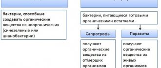
Prokaryotes (Greek πρό - before and κάρυον - nucleus) or prenuclear - single-celled organisms that, unlike eukaryotes, do not have a formed nucleus and membrane organelles. In prokaryotes, only non-membrane organelles can be found. Their genetic material is presented in the form of a circular DNA molecule - a nucleoid. Prokaryotes include bacteria (including cyanobacteria) and archaea.
Eukaryotes (Greek εὖ - good + κάρυον - nucleus) or nuclear - the domain of living organisms, the cells of which contain a formed nucleus. Plants, animals, fungi are eukaryotes.
Non-membrane organelles
- Ribosome
- Microtubules and microfilaments
- Cellular center (centrosome, from Greek soma - body)
- Cilia and flagella
A very small organelle (about 20 nm), which was discovered after the advent of the electron microscope. It consists of two subunits: large and small, which include proteins and rRNA (ribosomal RNA), synthesized in the nucleolus.
Remember the association: “The ribosome is a protein factory.” It is here that, during matrix biosynthesis - translation, which we will get to know in more detail in the following articles, a protein is synthesized on the basis of mRNA (messenger RNA) - a sequence of connected amino acids in the order specified by the mRNA.
Microtubules are intracellular protein derivatives that are part of the cytoskeleton. They maintain a certain cell shape and participate in the process of division by forming spindle threads. Microtubules also form the basis of the organelles of movement: flagella and cilia.
Microfilaments are thin, long thread-like structures made of the protein actin. They are found throughout the cytoplasm, serve to create a cytoplasmic current, and take part in cell movement and in the processes of endo- and exocytosis.
This organelle is characteristic only of animal cells; it is absent in the cells of fungi and higher plants. The cell center consists of 9 triplets of microtubules (a triplet is three connected together). Participates in the formation of spindle filaments and is located at the poles of the cell.
These are movement organelles that protrude above the cell surface and are based on a bundle of microtubules. Cilia are found only in animal cells; flagella can be found in animals, plants and bacteria.
Single-membrane organelles
- Endoplasmic reticulum (ER), endoplasmic reticulum (lat. reticulum - network)
- Golgi complex (apparatus)
- Lysosome (Greek lisis - dissolution + soma - body)
- Peroxisomes (Latin per - over, Greek oxys - sour and soma - body)
- Vacuoles
The ER is a system of membranes that permeate the entire cell and divide it into separate isolated parts (compartments). This is extremely important, since reactions take place in different parts of the cell that can interfere with each other, which will disrupt vital processes.
There are smooth EPS and rough EPS. Both of them perform the function of intracellular transport of substances, but there are differences between them. On the membranes of the smooth EPS, lipids are synthesized and harmful substances are neutralized. Rough ER synthesizes protein, as it has numerous ribosomes on its membranes (that is why it is called rough).
The Golgi complex consists of tubules, a network of flattened tubules (cisternae) and associated vesicles. Located around the cell nucleus, it looks like a stack of pancakes. This is a “cellular warehouse”. It stores fats and carbohydrates, with which chemical modifications occur here.
Modified substances are packaged in vesicles and can move to the cell membrane, connecting with it, they pour out their contents into the external environment. You can guess that the Golgi complex is well developed in the cells of the endocrine glands, which synthesize and release hormones in large quantities into the blood.
In the Golgi complex, primary lysosomes appear, which contain enzymes in an inactive state.
It is a membrane vesicle containing enzymes inside - lipases, proteases, phosphatases. The lysosome can be associated with a “cellular stomach”.
The lysosome is involved in the intracellular digestion of substances entering the cell. Merging with the phagosome, the primary lysosome turns into a secondary one, and the enzymes are activated. After the breakdown of substances, a residual body is formed - a secondary lysosome with undigested residues, which are removed from the cell.
The lysosome can digest the contents of the phagosome (the most harmless), digest part of the cell or the entire cell. Normally, each cell's life cycle ends with apoptosis, a programmed process of cell death.
During apoptosis, lysosome enzymes are released into the cell and its contents are digested. It is believed that disruption of apoptosis in cancer cells leads to uncontrolled tumor growth.
Peroxisomes (microbodies) contain redox enzymes that break down H2O2 (hydrogen peroxide) into water and oxygen. If hydrogen peroxide were left undegraded, it would cause severe cell damage.
Vacuoles are characteristic of plant cells, but are also found in animals (in unicellular organisms there are contractile vacuoles). In plants, vacuoles perform other functions and have a different structure: they are filled with cell sap, which contains a supply of nutrients. The outside of the vacuole is surrounded by tonoplast.
It is difficult to overestimate the importance of vacuoles in the life of a plant cell. Vacuoles create osmotic pressure and give the cell its shape.
It is noteworthy that the size of the vacuoles can be used to judge the age of the cell: young cells have small vacuoles, while in old cells the vacuoles can become so enlarged that they push the nucleus and other organelles to the periphery.
Double membrane organelles
- Core (“core” in Latin - nucleus, in Greek - karyon)
- Mitochondria
- Plastids (ancient Greek πλαστός - fashioned)
- Chloroplast (Greek chlōros - green)
- Chromoplasts (Greek chromos - paint)
- Leukoplasts (ancient Greek λευκός - white)
The most important component of a eukaryotic cell is the formed nucleus, which is absent in prokaryotes. The inner part of the nucleus is represented by karyoplasm, in which chromatin is located - a complex of DNA, RNA and proteins, and one or more nucleoli.
The nucleolus is a place in the nucleus where the process of matrix biosynthesis - transcription - is actively taking place, which we will get to know in more detail in the following articles. During the day, observing the same cell, you can see a different number of nucleoli or not find any.
The nuclear envelope consists of two membranes and is penetrated by a large number of nuclear pores, through which communication occurs between the karyoplasm and the cytoplasm. The main functions of the nucleus are storage, protection and transmission of hereditary material to daughter cells.
Note that chromosomes are visible only at the moment of cell division. Chromosomes are highly coiled DNA molecules bound to proteins.
I always recommend that students associate a chromosome with a skein of thread: if all the threads are wound around one axis, then they become a skein and are clearly visible (chromosomes - during division, spiralized DNA), but if the cell does not divide, then the threads are unwound and scattered in one layer , chromosomes are not visible (chromatin is decoiled DNA).
Chromosomes differ from each other in structure, shape, and size. The set of all characteristics (shape, number, size) of chromosomes is called a karyotype. A karyotype can be presented in different ways: there is a karyotype of a species, an individual, a cell.
By studying a person's karyotype, a geneticist can detect various hereditary diseases, for example, Down syndrome - trisomy on the 21st pair of chromosomes (there should be 2 chromosomes, but with Down syndrome there are three).
Rod-shaped organoid. Mitochondria can be compared to an “energy station.” If the anaerobic stage of respiration occurs in the cytoplasm (oxygen-free), then a more advanced stage occurs in the mitochondria - the aerobic stage (oxygen-free). As a result of the oxygen stage (Krebs cycle), 36 ATP molecules are obtained from two molecules of pyruvic acid (formed from 1 glucose).
The mitochondrion is surrounded by two membranes. Its inner membrane forms protrusions inward - cristae, on which there is a large accumulation of oxidative enzymes involved in the oxygen stage of respiration. Inside the mitochondrion is filled with a matrix.
Remember that a feature of this organelle is the presence of a circular DNA molecule - a nucleoid, and ribosomes. That is, the mitochondrion has its own genetic material and the ability to synthesize protein, almost like a separate organism.
In this regard, the mitochondrion is considered a semi-autonomous organelle. Most likely, mitochondria were initially independent organisms, but over time they entered into symbiosis with eukaryotes and became part of the cell.
Mitochondria are especially abundant in muscle cells, including cardiac muscle tissue. These cells perform active work and need a lot of energy.
Double-membrane organelles found only in the cells of higher plants, algae and some protozoa. The vast majority of animals do not have plastids. Divided into three types:
It got its name due to the green pigment it contains - chlorophyll (Greek chloros - green and phyllon - leaf). Under the double membrane are thylakoids, which are collected in stacks - grana. The inner space between the thylakoids and the membrane is called the stroma.
Remember that the light-dependent (light) phase of photosynthesis occurs on the thylakoid membranes, and the dark (light-independent) phase occurs in the stroma of the chloroplast due to the Calvin cycle. This will be very useful when studying photosynthesis in the future.
Just like mitochondria, plastids are semi-autonomous organelles: they contain circular DNA - a nucleoid, and ribosomes.
Plastids that contain carotenoid pigments in various combinations. The combination of pigments produces red, orange or yellow coloration. Found in fruits, leaves, flower petals.
Chromoplasts can develop from chloroplasts: during fruit ripening, chloroplasts lose chlorophyll and starch, and the biosynthesis of carotenoids is activated in them.
They do not contain pigments and are formed in the storage parts of the plant (tubers, rhizomes). Leucoplasts accumulate starch, lipids (fats), and peptides (proteins). In the light, leucoplasts can turn into chloroplasts and start the process of photosynthesis.
© Bellevich Yuri Sergeevich 2018-2020
This article was written by Yuri Sergeevich Bellevich and is his intellectual property. Copying, distribution (including by copying to other sites and resources on the Internet) or any other use of information and objects without the prior consent of the copyright holder is punishable by law. To obtain article materials and permission to use them, please contact Yuri Bellevich
.